The Translational Pathology Shared Resource is comprised of the Experimental Pathology (ExPath) Core and the Tissue Biorepository and Procurement Service (TBAPS). The UAMS ExPath Core Laboratory provides researchers with centralized, comprehensive histological services. The UAMS TBAPS is a repository of human specimens accessible to the UAMS research community.
Contacts
Director of the Translational Pathology Shared Resource
Behjatolah Karbassi, Ph.D.
501-526-7874
KarbassiBehjatolahM@uams.edu
ExPath Core
Jennifer James, ExPath Core Laboratory Manager
JamesJenniferD@uams.edu
TBAPS
Rémelle Eggerson, TBAPS Project Manager
EggersonRemelleM@uams.edu
Overview
The ExPath Core can process and paraffin embed tissues and section frozen and paraffin-embedded tissues for use in biochemical, molecular, and pathologic studies. The laboratory works with investigators to develop immunostaining approaches to detect specific targets of interest, and to identify clinical pathologists who can assist with image analysis and data interpretation. TBAPS uses an IRB approved protocol and general consent form to consent patients, collect samples from the pathology grossing room, and provide samples to researchers who have appropriate approval. Solid tissues and fluid specimens are stored in liquid nitrogen tanks. Solid tissues may also be embedded into paraffin. De-identified samples and related pathology data are available for a variety of organs.
Services
Experimental Pathology Core Laboratory
- Tissue processing and embedding
- Tissue sectioning (frozen and paraffin-embedded)
- Tissue staining (H&E, immunohistochemical, immunofluorescent, and special stains)
- Digital slide scanning
- Multispectral imaging and analysis
- Pathology consultation
Tissue Biorepository and Procurement Service
- Collection of solid tissue specimens (fresh, snap-frozen, and/or paraffin-embedded) and fluid specimens (serum and urine)
- Project specific tissue procurement and storage
- Assist with patient consenting
- Consult on protocol development for tissue acquisition
- Specimen management using secure, web-based data management software (OpenSource)
Equipment
Experimental Pathology
Processing: Tissue-Tek VIP 6 A1 Tissue Processor
Embedding: Tissue-Tek TEC 6 Embedding Center
Sectioning: Leica CM3050S Cryostat, Microm HM325 Rotary Microtome
Staining: Histo-Tek SL Automated Slide Stainer, Leica CV5030 Glass Coverslipper, Leica Bond RX Automated Research Stainer
Imaging: Digital imaging and analysis of slides are available using an Aperio CS2 Scanscope coupled with ImageScope Slide Viewer and Image Analysis software from Leica. Available analysis algorithms include color deconvolution, nuclear and membrane localization, and quantifying positive pixel number and intensity.
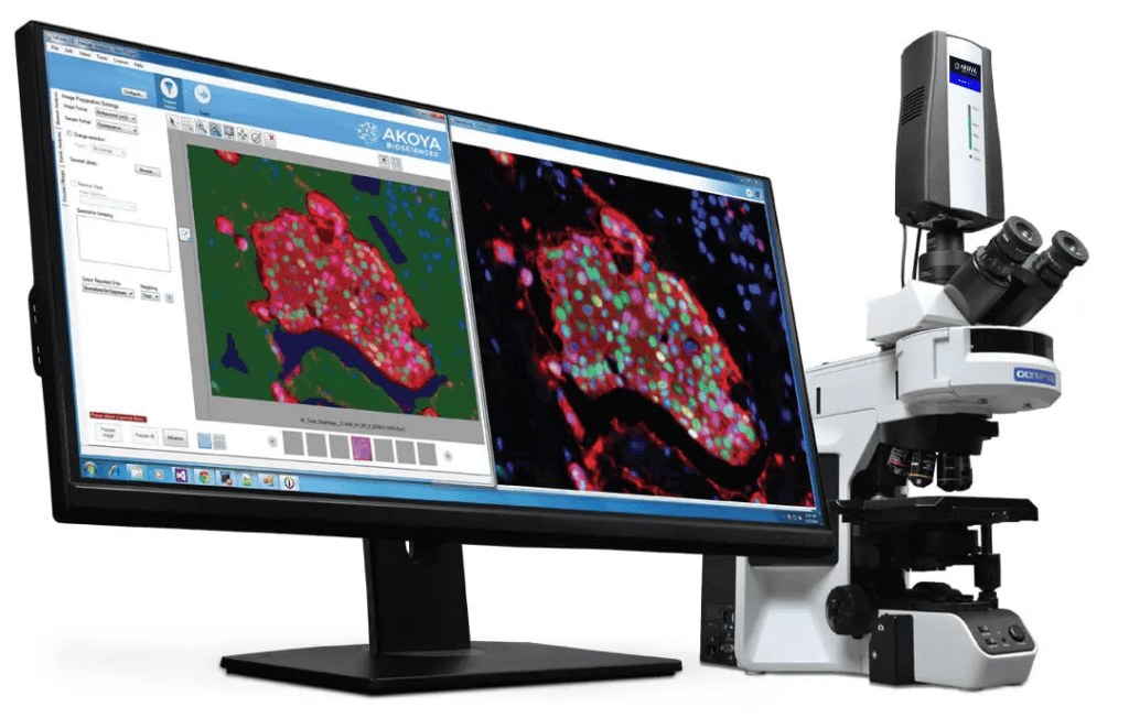
Phenoptics™ analysis is available using the Mantra™ Quantitative Pathology Workstation, which is a manual microscope equipped with a multispectral camera and inForm® image analysis software. Phenoptics integrates multiplexed immunohistochemistry and imaging to quantitatively capture systems biology data with cellular detail. It reveals multi-parameter cellular expressions and interactions while retaining spatial context, offering insights into the complexity of tissue biology.
Tissue Biorepository and Procurement
- Over 8,000 patients consented
- Over 20,000 samples banked
- 11 liqN2 freezers
- 15 -80 ℃ freezers
- 2 -20 ℃ freezers
- Table-top BD Accuri flow-cytometer
- 2 RoboSep automated cell separators
- Equipment for sample processing
- Domino/MAPI straw system for aliquoting liquid samples
- OpenSpecimen biobanking/biorepository management software